Electrodiagnostic Testing and its Relation to Eye Care

Electrodiagnostic testing measures the amount of electrical activity in certain areas of the body. Visual electrodiagnostics are used to measure the function of the patient’s retina and optic nerve and can diagnose certain disorders of the retina and brain. In order to measure the electrical activity, electrodes are attached to the patient’s skin on the head or near the eyes and is non-invasive and painless.

Electrodiagnostic Testing

When vision occurs, an image is projected onto the patient’s retina located at the back portion of the eye. The retina’s visual function is to convert the projected image into tiny electrical signals which are then passed to the brain via the optic nerve. As a result, the patient can then see the image. Visual electrodiagnostic testing works by measuring the electrical signals within the eye and brain.

By examining the signals, eye care professionals can assess the patient’s level of retinal and optic nerve function. If abnormalities in the electrical signals are identified, the eye care professional can investigate further to determine if the patient suffers certain brain or retina disorders.

Electrodiagnostics Uses

Electrogiagnostic testing is typically used to examine the functioning of muscles and nerves in various parts of the body and can determine if a nerve is pinched or functioning abnormally. Electrodiagnostics can be used to evaluate the cause for a patient’s numbness, pain, weakness, muscle cramping, and fatigue.

In optometry, electrodiagnostics can help to identify ocular diseases such as retinal degeneration. Electrodiagnostics can also help determine retinal ischemic disorders, which are characterized by reduced blood flow to the patient’s eye and diagnose glaucoma, which results from damage to the optic nerve If left untreated, glaucoma can lead to vision damage or complete vision loss. Electrodiagnostics aid eye care professionals in diagnosing neurologic disorders as well.

Electroretinography

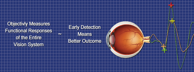
Electroretinography (ERG) is one of the most common forms of visual electrodiagnostic testing. ERG testing for the retina is similar to an electrocardiogram (ECG) test for the heart and uses flashes of light in both a lit and dark room. It records the electrical signals sent from the retina during this testing.

ERG testing examines both the rods and cones of the eye. The rods are responsible for night vision, and the cones are responsible for day vision. ERG utilizes varying flash rate, brightness, and light color to adequately assess the rods and cones separately. ERG testing can be helpful in diagnosing hereditary eye diseases such as retinitis pigmentosa, a disease that can cause severe vision impairment and blindness.

Visual Evoked Response

Visual Evoked Response (VER) testing is used to investigate the pathways that carry signals from the patient’s eye to the brain. VER also examines the optic nerves as well as the process through which the brain interprets these visual signals.

During VER testing, the patient is typically asked to observe flashes of light or a moving pattern such as a checkerboard. The eye care professional can then record the electrical signals that are being communicated within the eye and brain. Using VER testing, eye care professionals can determine damage to visual pathways.