 Computer-assisted videokeratography (CAVK) devices create a three-dimensional map of the patient’s cornea and is a simple and completely painless technique for evaluating corneal health. During a CVAK procedure, the patient is seated in front of a bowl that contains an illuminated pattern on a Placido cone disk. This pattern is typically a layering of concentric rings.
The eye care professional focuses the pattern onto the cornea’s anterior surface. The pattern is then reflected into a digital camera that is located at the center of the Placido cone, which is named after 1880 Portuguese ophthalmologist, Antonio Placido. He discovered the basic concept of reflecting rings onto the cornea to reveal the cornea’s contour lines.
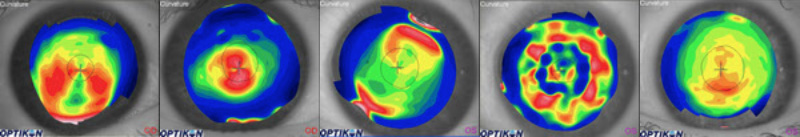
The shape taken by the device’s reflected pattern reveals the cornea’s topography. Within seconds, the CAVK computer analyzes the data. This data provides the eye care professional with a grid containing thousands of points in varying heights and positions across the cornea. With this information, topographical maps can be created for evaluation by the eye care professional. Topographical maps can be displayed through several graphical formats. A sagittal map, for example, is color-coded according to the steepness of the cornea’s curvature.
 The Keratron is a standard of reference in corneal topography technology and offers exceptional precision and test repeatability. The Keratron is able to highlight the most subtle corneal details and uses a proprietary camera and software to digitally map the cornea with thousands of numbers. The doctor utilizes this mapped data and computer aided design software to evaluate and treat various corneal conditions.

The Keratron features a patented Eye Position Control System (EPCS) to capture corneal images only at the correct intended focusing distance. The system automatically corrects misalignments to ensure accuracy. The Keratron displays the patient’s tear film and still images on a large, high-resolution monitor.
The device features a Placido cone with 28 rings. The Keratron’s cone allows up to 90 percent of the corneal surface to be analyzed. The device is utilized to analyze refractive surgery candidates, diagnose corneal disease, and design custom contact lenses.