Fundus Autofluorescence Imaging

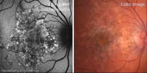
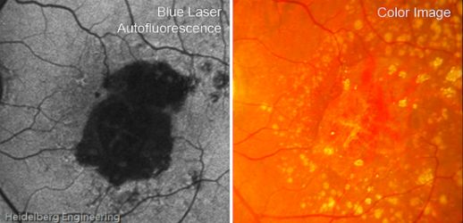
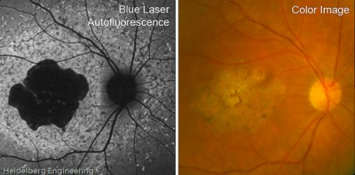
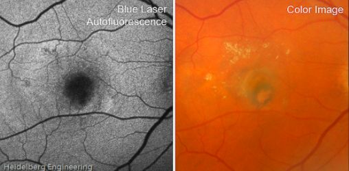
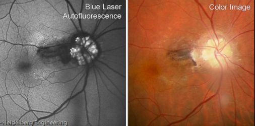
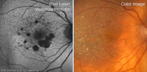
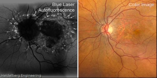
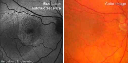
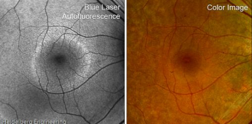
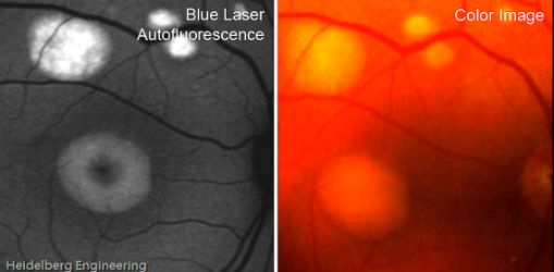
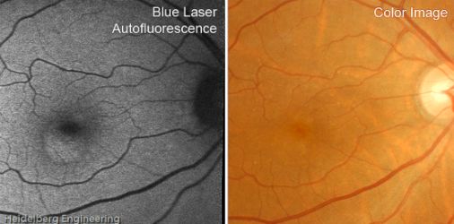
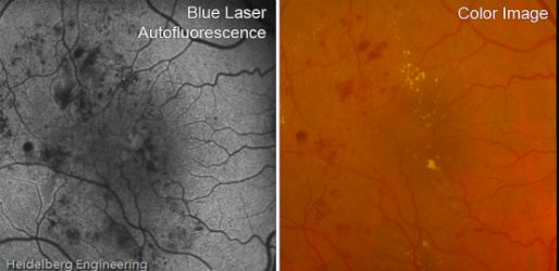
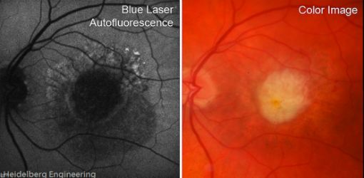
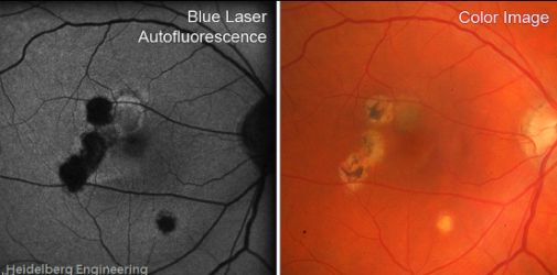
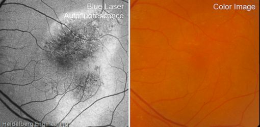
Fundus autofluorescence (FAF) imaging is an advanced diagnostic technique for observing the fundus. It is a noninvasive technique, making it a relatively simple and effective method for diagnosing AMD. Fundus autofluorescence detects chemical structures called fluorophores. When exposed to a particular wavelength of light, fluorophores become fluorescent, or illuminated.
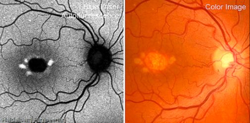
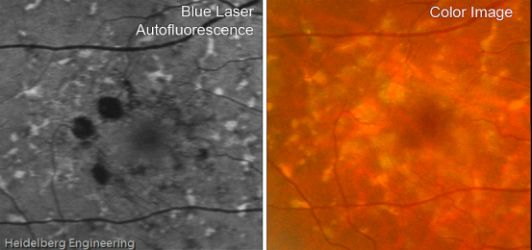
What Is a Fundus?:

A fundus is defined as the part of an organ that is opposite the organ’s opening. The fundus of the eye is the eye’s interior surface, opposite the lens. The fundus includes the retina, macula and fovea, optic disc, and posterior pole. Eye care professionals often observe the fundus through special imaging and technology which can detect eye disease such as age-related macular degeneration (AMD), hemorrhages, and blood vessel abnormalities.

Detection of Lipofuscin

Fundus autofluorescence imaging pays particular attention to a fluorescent pigment lipofuscin. This pigment is a metabolic waste product that can be found in many organs. In the eye, lipoduscin is a byproduct of the incomplete degradation of the outer segments of photoreceptor cells.

Typically, lipofuscin increases in the retinal pigment epithelium (RPE) of the eye as a patient becomes older. However, high levels of lipofuscin can also be caused by cell dysfunction that can be attributed to eye diseases such as age-related macular degeneration (AMD). The fluorophore in lipofuscin has toxic properties and can interfere with cell function within the eyes.

Fundus Autofluorescence for AMD Diagnosis

Fundus autofluorescence is believed to benefit patients who have been diagnosed with either dry or wet AMD, called geographic atrophy and choroidal neovascularization. The dry form is known as central geographic atrophy (GA). This atrophy is a type of degeneration that reduces the size of a tissue layer below the retina which results in a reduction in the rods and cones (photoreceptors) in the center of the eye.

Choroidal neovascularization is a wet form of AMD. The wet form of AMD causes vision loss by affecting blood vessel growth which eventually leads to protein and blood leakage around the macula. If the leakage results in untreated scarring, irreversible vision loss can result.